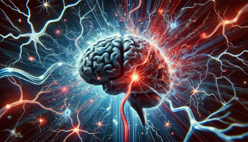
The Significance of Imaging in Brain Trauma Diagnosis
The human brain is one of the most intricate and vital organs, governing every aspect of thought, emotion, and movement. When brain trauma occurs, the ability to assess the extent of the damage becomes crucial for effective treatment and rehabilitation. Imaging techniques, particularly head trauma MRI and traumatic brain injury CT scans, play a critical role in identifying structural and functional impairments. These advanced diagnostic tools provide insights into how traumatic events disrupt neural pathways, potentially leading to cognitive and motor deficits. Understanding the mechanisms of MRI brain damage detection allows both medical professionals and individuals affected by brain trauma to develop informed treatment strategies that optimize recovery outcomes.
You may also like: How to Retrain Your Brain: Harnessing Neuroplasticity to Overcome Anxiety and Strengthen Neural Pathways
The Mechanisms Behind Brain Trauma and Neural Pathway Disruption
Brain trauma can occur due to various incidents, including vehicular accidents, falls, sports injuries, and violent impacts. When the brain experiences a sudden jolt or collision, the delicate neurons and synapses within its structure may sustain irreversible damage. Depending on the severity, this can range from minor concussions to severe cases of hemorrhaging, swelling, and axonal shearing. In many cases, standard imaging techniques like a traumatic brain injury CT scan can detect immediate issues such as skull fractures and internal bleeding. However, a head trauma MRI is often necessary to evaluate the more intricate details of neural integrity, as it provides a clearer picture of microstructural damage that may not be visible on other imaging modalities.
How MRI and CT Scans Differ in Diagnosing Traumatic Brain Injuries
Both MRI and CT scans are widely used in diagnosing traumatic brain injuries, but they serve different purposes. A traumatic brain injury CT scan is typically the first step in emergency settings, as it provides a rapid assessment of acute injuries, such as bleeding, fractures, and swelling. CT scans use X-ray technology to generate detailed cross-sectional images, making them essential for identifying life-threatening conditions that require immediate intervention. However, when it comes to detecting subtle neural disruptions and long-term effects, MRI is the superior choice. Head trauma MRI scans utilize magnetic fields and radio waves to create high-resolution images of the brain’s soft tissues, revealing signs of diffuse axonal injury, microhemorrhages, and white matter degradation. The enhanced imaging capabilities of brain trauma MRI make it an indispensable tool for assessing long-term neurological impairments and guiding rehabilitation strategies.
Neural Pathway Damage: Short-Term and Long-Term Implications
When the brain sustains trauma, the immediate impact on neural pathways can result in symptoms ranging from mild confusion to complete loss of consciousness. Short-term impairments often include headaches, dizziness, difficulty concentrating, and mood disturbances. In more severe cases, damaged neural pathways can lead to long-term cognitive decline, memory loss, and motor dysfunction. MRI brain damage assessments help identify whether the injury has caused persistent disruptions in communication between different brain regions. This is particularly important for individuals experiencing post-concussive symptoms, as lingering damage to neural circuits can affect daily functioning. With the help of advanced imaging, clinicians can pinpoint the affected areas and develop targeted therapies aimed at restoring optimal neural connectivity.
The Role of MRI in Monitoring Brain Recovery Post-Trauma
A significant advantage of head trauma MRI is its ability to track changes in brain structure over time. Unlike static imaging techniques, MRI allows medical professionals to monitor the healing process and assess whether neural pathways are recovering or deteriorating. Repeated MRI scans over weeks or months provide valuable insights into how the brain compensates for lost function through neuroplasticity. In cases where rehabilitation efforts are not yielding the expected improvements, imaging data can help refine treatment plans, ensuring that patients receive the most effective interventions. This underscores the importance of utilizing MRI not only as a diagnostic tool but also as a means of evaluating recovery trajectories.
How Neuroplasticity Contributes to Healing After Brain Trauma
Despite the significant impact of brain injuries, the human brain possesses an extraordinary ability to adapt and reorganize itself. This phenomenon, known as neuroplasticity, plays a crucial role in recovery following neural damage. When a traumatic injury disrupts normal brain function, undamaged neurons can form new connections to compensate for lost pathways. Rehabilitation programs focusing on cognitive exercises, physical therapy, and sensory stimulation help facilitate this process, encouraging the brain to rewire itself. Brain trauma MRI studies have demonstrated that patients who actively engage in targeted therapies show increased neural connectivity in previously affected areas. This highlights the importance of structured rehabilitation in maximizing the brain’s capacity for recovery and reducing long-term deficits.
Emerging Technologies and Future Directions in Brain Trauma Diagnosis
Recent advancements in imaging technology continue to enhance our understanding of traumatic brain injuries and their long-term effects. Researchers are developing more sophisticated MRI techniques, such as functional MRI (fMRI) and diffusion tensor imaging (DTI), which provide even deeper insights into neural pathway disruptions. fMRI measures brain activity by detecting changes in blood flow, allowing scientists to assess how different regions of the brain communicate post-injury. Meanwhile, DTI specializes in mapping white matter tracts, revealing patterns of axonal damage that traditional scans might miss. These innovations are paving the way for more accurate diagnoses and personalized treatment plans tailored to each patient’s unique neural profile.
The Importance of Early Detection and Preventative Measures
While head trauma MRI and traumatic brain injury CT scans are invaluable for diagnosing and managing brain injuries, prevention remains the best approach. Wearing protective gear during high-risk activities, practicing safe driving habits, and addressing health conditions that increase fall risk can significantly reduce the likelihood of head trauma. Additionally, recognizing the early signs of brain injuries—such as persistent headaches, memory problems, and changes in mood—ensures that individuals seek medical attention before symptoms worsen. By prioritizing both prevention and early intervention, the long-term effects of brain injuries can be mitigated, improving overall cognitive health and quality of life.
Frequently Asked Questions (FAQ) About Brain Trauma and Imaging
1. What advantages does a traumatic brain injury MRI have over other imaging techniques?
A traumatic brain injury MRI offers superior visualization of soft tissue structures, allowing for detailed examination of neural damage that may not be visible on other scans. Unlike a traumatic brain injury CT scan, which is primarily used for detecting acute bleeding and skull fractures, MRI provides a clearer picture of microstructural injuries, such as diffuse axonal injury and white matter degradation. This precision makes MRI invaluable for assessing long-term cognitive and neurological effects following brain trauma. Additionally, newer MRI techniques, such as functional MRI (fMRI) and diffusion tensor imaging (DTI), offer insights into brain activity and neural connectivity, helping doctors design more effective rehabilitation plans. Given its detailed imaging capabilities, head trauma MRI is often preferred for evaluating complex cases where cognitive symptoms persist without obvious structural abnormalities.
2. Can an MRI detect brain damage that a CT scan might miss?
Yes, an MRI is often more effective than a traumatic brain injury CT scan when detecting subtle or long-term brain damage. While CT scans are excellent for identifying acute injuries like fractures and hematomas, they may not detect smaller lesions, microscopic hemorrhages, or shearing injuries affecting neural pathways. MRI brain damage assessments can reveal disruptions in brain function that a CT scan might overlook, making it a preferred method for evaluating patients with lingering cognitive or neurological deficits. Furthermore, MRI scans provide better contrast resolution, allowing clinicians to assess even minor changes in brain tissue over time. This makes brain trauma MRI an essential tool for tracking recovery and guiding long-term treatment strategies.
3. How does brain trauma MRI contribute to personalized rehabilitation programs?
MRI imaging not only helps diagnose brain trauma but also plays a crucial role in shaping individualized rehabilitation plans. By mapping neural damage and identifying affected regions, doctors can tailor cognitive therapy, physical rehabilitation, and neurofeedback treatments to each patient’s unique condition. For example, if an MRI brain damage scan reveals disruptions in the prefrontal cortex, treatment might focus on executive functioning and decision-making exercises. Likewise, if white matter damage is detected, therapies aimed at restoring neural communication pathways can be prioritized. This level of precision ensures that rehabilitation efforts align with the specific nature of the brain injury, leading to more effective recovery outcomes.
4. Are there limitations to using MRI for diagnosing traumatic brain injuries?
While head trauma MRI is a powerful diagnostic tool, it does have some limitations. One of the biggest challenges is accessibility, as MRI machines are more expensive and less readily available than CT scanners, particularly in emergency settings. Additionally, MRI scans require more time to perform, which may not be ideal for patients who need immediate medical intervention. Some patients, particularly those with metal implants or claustrophobia, may not be suitable candidates for MRI. Moreover, although MRI brain damage assessments provide detailed images, interpreting these results requires a high level of expertise to distinguish between pre-existing conditions and trauma-related abnormalities. Despite these limitations, MRI remains the gold standard for evaluating complex or persistent brain injuries.
5. How does repeated imaging help monitor recovery from a traumatic brain injury?
Traumatic brain injury MRI scans are often performed at multiple intervals to track a patient’s recovery over time. This allows doctors to assess how well damaged brain tissue is healing and whether neural pathways are reestablishing connectivity. If changes in brain structure indicate improvement, rehabilitation programs can be adjusted to reinforce positive gains. Conversely, if an MRI reveals continued deterioration or the presence of new abnormalities, treatment plans can be modified to address these issues promptly. Repeated imaging also provides critical insights for research into how different rehabilitation strategies influence brain plasticity and recovery rates, ultimately benefiting future patients with similar conditions.
6. Can brain trauma MRI predict long-term cognitive impairment?
Yes, brain trauma MRI scans can provide valuable insights into the likelihood of long-term cognitive impairment. By analyzing the extent and location of neural damage, clinicians can make informed predictions about potential challenges a patient may face. For example, injuries affecting the hippocampus might indicate future memory difficulties, while damage to the frontal lobe could impact decision-making and emotional regulation. Early detection of these vulnerabilities allows for proactive intervention, including targeted cognitive therapy and lifestyle modifications. Although MRI cannot provide absolute certainty regarding long-term outcomes, it serves as a crucial tool for early intervention and personalized treatment planning.
7. How do imaging advancements improve the accuracy of brain injury diagnosis?
Recent advancements in imaging technology have significantly enhanced the accuracy of brain trauma assessments. Techniques such as diffusion tensor imaging (DTI) allow for precise mapping of white matter pathways, helping to identify even the most subtle disruptions. Functional MRI (fMRI) measures blood flow and neural activity, providing a real-time view of brain function after an injury. Additionally, artificial intelligence (AI) is being integrated into imaging analysis to detect patterns in traumatic brain injury MRI scans that may be overlooked by the human eye. These innovations are improving diagnostic precision and leading to more effective, data-driven treatment strategies. As technology evolves, the ability to detect and manage brain injuries will continue to improve, offering hope for better recovery outcomes.
8. What role does early imaging play in preventing long-term complications?
Early imaging with a traumatic brain injury CT scan or MRI is critical for preventing long-term complications. Identifying brain damage soon after an injury allows for timely medical interventions that can minimize secondary complications such as swelling, hemorrhaging, or increased intracranial pressure. Additionally, early detection of neural pathway disruptions can guide immediate therapeutic interventions, reducing the risk of chronic cognitive deficits. The sooner an injury is diagnosed and managed, the better the chances of optimizing neuroplasticity and functional recovery. Delayed diagnosis can result in missed opportunities for treatment, potentially leading to irreversible impairments that affect an individual’s quality of life.
9. Can lifestyle changes complement MRI-guided brain trauma recovery?
Yes, lifestyle changes can play a significant role in supporting recovery from MRI-detected brain trauma. Nutritional strategies, including a diet rich in omega-3 fatty acids and antioxidants, help protect neurons and promote repair. Regular physical activity enhances cerebral blood flow, aiding in neuroplasticity and cognitive function. Mindfulness practices and stress reduction techniques help regulate the nervous system and prevent further neural strain. Sleep hygiene is also crucial, as deep sleep supports memory consolidation and synaptic repair. Combining these lifestyle adjustments with MRI-guided rehabilitation strategies maximizes the brain’s potential for healing and cognitive restoration.
10. How can patients and caregivers advocate for advanced imaging in brain trauma cases?
Patients and caregivers can play a proactive role in ensuring that advanced imaging, such as head trauma MRI, is included in the diagnostic process. If symptoms persist despite an initial traumatic brain injury CT scan showing no major abnormalities, advocating for an MRI can provide deeper insights into potential microstructural damage. Educating oneself about the benefits of MRI and discussing persistent cognitive or neurological issues with a healthcare provider can strengthen the case for further imaging. Additionally, seeking second opinions from specialists in neurotrauma or neurology can help determine whether advanced imaging is necessary. Ensuring access to high-quality diagnostic tools is crucial for obtaining the most accurate assessment and tailoring an effective recovery plan.
Conclusion: The Power of Imaging in Understanding and Treating Brain Trauma
The ability to visualize brain injuries through advanced imaging techniques has revolutionized the field of neuroscience and trauma care. Head trauma MRI, along with complementary imaging methods like traumatic brain injury CT scans, provides an in-depth understanding of how neural pathways are affected by trauma. These tools not only aid in diagnosis but also guide rehabilitation efforts, enabling patients to regain cognitive function and quality of life. As research continues to push the boundaries of neuroimaging, the future holds promising possibilities for improving both the accuracy of brain trauma diagnosis and the effectiveness of recovery strategies. Understanding the role of MRI brain damage assessment in detecting and treating traumatic injuries empowers individuals to take proactive steps toward brain health, ensuring resilience against the long-term effects of neural disruption.
Further Reading:
Neuroimaging in Traumatic Brain Imaging
Imaging for the Diagnosis and Management of Traumatic Brain Injury
Important Note: The information contained in this article is for general informational purposes only, and should not be construed as health or medical advice, nor is it intended to diagnose, prevent, treat, or cure any disease or health condition. Before embarking on any diet, fitness regimen, or program of nutritional supplementation, it is advisable to consult your healthcare professional in order to determine its safety and probable efficacy in terms of your individual state of health.
Regarding Nutritional Supplements Or Other Non-Prescription Health Products: If any nutritional supplements or other non-prescription health products are mentioned in the foregoing article, any claims or statements made about them have not been evaluated by the U.S. Food and Drug Administration, and such nutritional supplements or other health products are not intended to diagnose, treat, cure, or prevent any disease.